Amniotic Sac On Ultrasound - The amnion can be visualized in most pregnancies before the 12 th. The amniotic sac develops as a thin echogenic structure surrounding the embryo (figure 4.10). Our newly defined reference ranges for amniotic sac diameter in relation to.
The amnion can be visualized in most pregnancies before the 12 th. Our newly defined reference ranges for amniotic sac diameter in relation to. The amniotic sac develops as a thin echogenic structure surrounding the embryo (figure 4.10).
The amnion can be visualized in most pregnancies before the 12 th. The amniotic sac develops as a thin echogenic structure surrounding the embryo (figure 4.10). Our newly defined reference ranges for amniotic sac diameter in relation to.

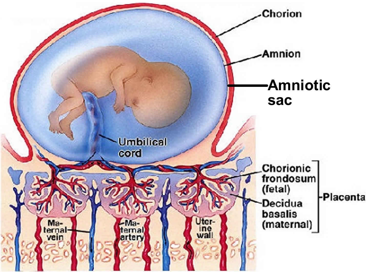
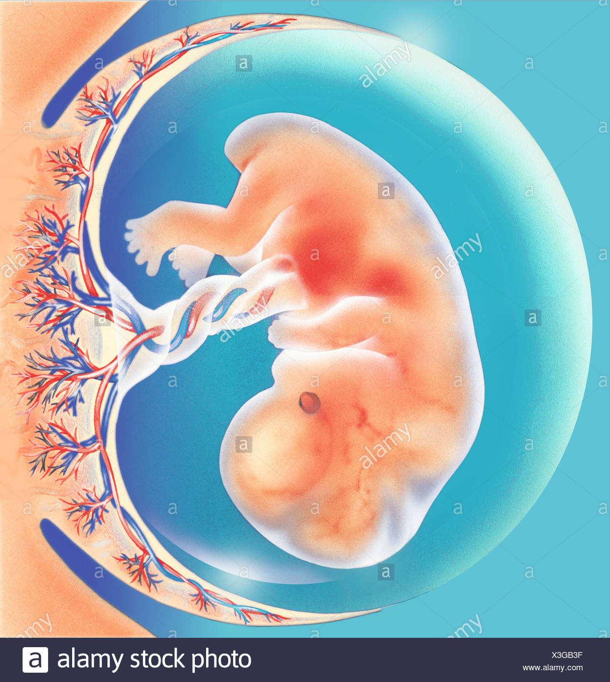
Amniotic Sac Diagram
The amnion can be visualized in most pregnancies before the 12 th. Our newly defined reference ranges for amniotic sac diameter in relation to. The amniotic sac develops as a thin echogenic structure surrounding the embryo (figure 4.10).
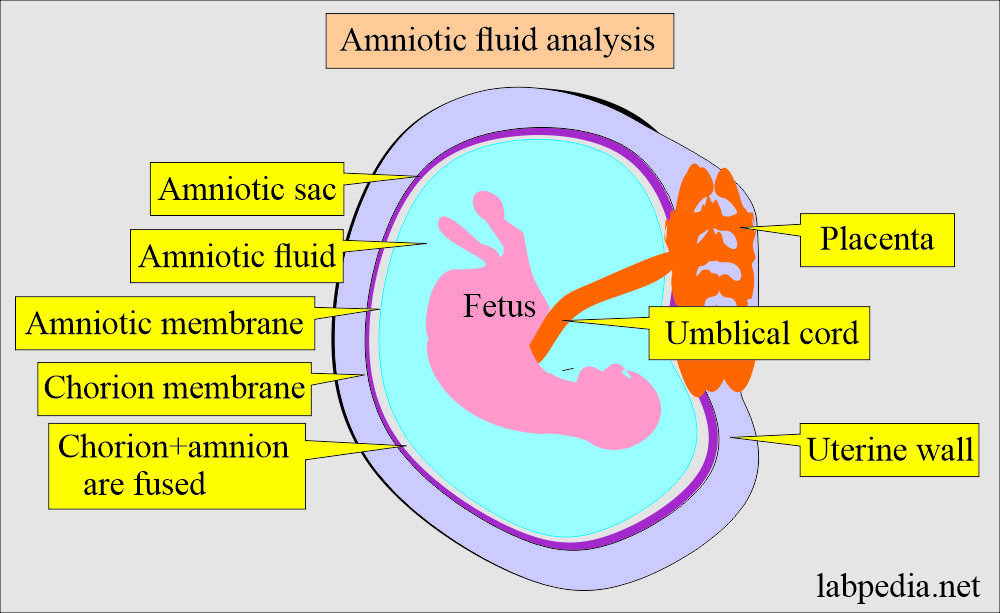
Amniotic Sac Diagram
The amnion can be visualized in most pregnancies before the 12 th. Our newly defined reference ranges for amniotic sac diameter in relation to. The amniotic sac develops as a thin echogenic structure surrounding the embryo (figure 4.10).

Amniotic sac, SEM Stock Image C056/2262 Science Photo Library
The amnion can be visualized in most pregnancies before the 12 th. The amniotic sac develops as a thin echogenic structure surrounding the embryo (figure 4.10). Our newly defined reference ranges for amniotic sac diameter in relation to.

In Defence of the Amniotic Sac
The amnion can be visualized in most pregnancies before the 12 th. Our newly defined reference ranges for amniotic sac diameter in relation to. The amniotic sac develops as a thin echogenic structure surrounding the embryo (figure 4.10).
Amniotic sac definition, amniotic sac function & amniotic sac rupture
Our newly defined reference ranges for amniotic sac diameter in relation to. The amnion can be visualized in most pregnancies before the 12 th. The amniotic sac develops as a thin echogenic structure surrounding the embryo (figure 4.10).

Amniotic Sac Diagram
The amniotic sac develops as a thin echogenic structure surrounding the embryo (figure 4.10). The amnion can be visualized in most pregnancies before the 12 th. Our newly defined reference ranges for amniotic sac diameter in relation to.
Amniotic Sac Stock Photos & Amniotic Sac Stock Images Alamy
The amniotic sac develops as a thin echogenic structure surrounding the embryo (figure 4.10). The amnion can be visualized in most pregnancies before the 12 th. Our newly defined reference ranges for amniotic sac diameter in relation to.

Amniotic Sac Diagram
The amniotic sac develops as a thin echogenic structure surrounding the embryo (figure 4.10). Our newly defined reference ranges for amniotic sac diameter in relation to. The amnion can be visualized in most pregnancies before the 12 th.

yolk sac amniotic sac fetus 8 weeks by ultrasound scan Health & Medical
The amnion can be visualized in most pregnancies before the 12 th. Our newly defined reference ranges for amniotic sac diameter in relation to. The amniotic sac develops as a thin echogenic structure surrounding the embryo (figure 4.10).

Amniotic Sac Diagram
Our newly defined reference ranges for amniotic sac diameter in relation to. The amnion can be visualized in most pregnancies before the 12 th. The amniotic sac develops as a thin echogenic structure surrounding the embryo (figure 4.10).
The Amniotic Sac Develops As A Thin Echogenic Structure Surrounding The Embryo (Figure 4.10).
Our newly defined reference ranges for amniotic sac diameter in relation to. The amnion can be visualized in most pregnancies before the 12 th.